Lab Tests & Radiology
Fluoroscopy (FLUORO)

What is fluoroscopy?
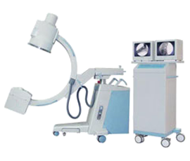
Fluoroscopy is a study of moving body structures. Think of it as an X-ray "movie." A continuous X-ray beam is passed through the body part being examined, and is transmitted to a monitor so that the body part and its motion can be seen in detail.
Fluoroscopy is an imaging tool enabling physicians to look at many body systems including:
- skeletal
- digestive
- urinary
- respiratory
- reproductive
Fluoroscopy may be performed to evaluate specific areas of the body, including the bones, muscles, and joints, as well as solid organs such as the heart, lung, or kidneys and is used in many types of examinations and procedures, such as:
- barium X-rays
- cardiac catheterization
- arthrography (visualization of a joint or joints)
- lumbar puncture
- placement of intravenous catheters (hollow tubes inserted into veins or arteries)
- intravenous pyelogram
- hysterosalpingogram
- biopsies.
As a diagnostic procedure, Fluoroscopy may be used alone or in conjunction with other diagnostic or therapeutic media or procedures.
In barium X-rays, fluoroscopy used alone allows the physician to see the movement of the intestines as the barium moves through them.
In cardiac catheterization, fluoroscopy is added to enable the physician to see the flow of blood through the coronary arteries in order to evaluate the presence of arterial blockages.
For intravenous catheter insertion, fluoroscopy assists the physician in guiding the catheter into a specific location inside the body.
Other uses of fluoroscopy include, but are not limited to:
- locating foreign bodies.
- viscosupplementation injections of the knees - a procedure in which a liquid substance that acts as a cartilage replacement or supplement is injected into the knee joint.
- image-guided anesthetic injections into joints or the spine.
- percutaneous vertebroplasty - a minimally invasive procedure used to treat compression fractures of the vertebrae of the spine.
How is fluoroscopy performed?
Fluoroscopy may be part of an examination or procedure that can done on an outpatient or inpatient basis. The specific type of procedure/examination being done will determine whether any preparation prior to the procedure is required. Your physician should notify you of any pre-procedure instructions.
Each facility may have specific protocols in place and specific examinations and procedures may differ.
How do I schedule a fluoroscopy procedure?
If your Physician has stated to you that you need to have a Fluoroscopy procedure, contact the Radiology Department at 337-531-3376. Monday - Friday, 8 a.m. - 4:30 p.m. to schedule your exam.
What is an Upper GI?
An upper GI is a series of X-rays examining the esophagus, stomach and first part of the small intestine (duodenum). These parts of the body are known as the upper gastrointestinal (GI) tract or upper digestive system.
A barium swallow is used when only the pharynx and esophagus are evaluated. In order for these parts to show up on an X-ray the upper gastrointestinal tract must be coated or filled with a contrast material called barium. Barium is an element that appears white on X-rays.
Patient Preparation for Upper GI:
Your physician will give you detailed instructions on how to prepare for your Upper GI imaging. Be sure that he or she is aware of all the medications you are taking. There may be some medications that you will need to stop taking before your exam. You will be asked not eat or drink anything four to eight hours prior to your exam.
The quality of the images obtained during the procedure can be affected if the stomach is not empty of food. You should also not chew gum or smoke after the specific time you are given as these activities can increase stomach secretions that may affect the quality of the images.
What you can expect for an Upper GI:
An upper GI is usually done in a hospital or imaging center on an outpatient basis. They are usually scheduled in the morning to reduce your time of fasting. When you arrive, you will be asked to change into a gown and remove all jewelry. You will be sitting or standing up while your heart, lungs and abdomen are examined with a fluoroscope. You may be asked to swallow baking soda crystals before you drink the contrast material. This helps create gas in your stomach and increase visibility.
The technologist will position you standing next to the X-ray equipment and then you will be asked to drink a cup of liquid barium, which resembles a light colored milkshake and has a chalky consistency. As you drink the barium, the Radiologist will note and monitor the passage of the liquid into your esophagus and stomach on the monitor. Once the upper gastrointestinal tract is adequately coated with the barium, X-rays will be taken.
In some cases, your physician may order a detailed intestine or small bowel follow through (SBFT). Once the preliminary upper GI series is complete, you will be escorted to a waiting area while the barium travels down through the rest of the small intestines. Every 15-30 minutes you will return to the X-ray suite for additional films. Once the barium has completed its trip down the small bowel tract, the test is complete. You may be in the department for this procedure for up to four hours.
After the exam:
When your test is complete, you may resume your regular diet and restart any medications you were asked to stop according to you physicians instructions. You should drink an extra four to eight glass of water after your exam to help move the barium through your system.
Your stools may appear gray or white up to 72 hours following this procedure. If you experience constipation, your physician may recommend a mild laxative.
What is a Lower GI:
The lower gastrointestinal (GI) examination or "Barium Enema" will be performed by a Radiologist specializing in G.I. examinations.
For this exam, you will change from your clothing into hospital clothing. The technologist will gently position you on a special tilting table attached to a Fluoroscope. As you lie on your side, a lubricated enema tube will be inserted into your rectum, and a liquid barium mixture will be released.
The tip of the enema tube is specially designed to help hold the barium, however, as the barium fills your colon, you may feel like you need to move your bowel. Let the technologist or the Radiologist know if you are having trouble holding the barium in.
Using the Fluoroscope, the Radiologist will observe the barium flow into your colon. You will be asked to turn from side to side and hold several different positions. During this time, some gentle pressure may be applied to your abdomen and the table may be tilted slightly. After a series of X-rays have been completed, you will be allowed to go to the bathroom to expel the barium.
You may be asked to come back for further X-rays of your empty colon.
How do I prepare for this exam?
You physician will provide you with instructions for preparing for the examination. Two or three days before the exam, you probably will be instructed to eat a "low-residue" diet (liquids, low-fat and finely crushed foods). You may be asked to drink only clear liquids the day before the exam and you will probably be asked to refrain from drinking or eating anything after midnight the night before the exam.
For the examination to be successful, your lower digestive tract must be completely empty. Any residue will show up on the X-rays and could be mistaken for an abnormality in your colon or rectum. Your physician may also request that you take a mild laxative or suppository to help clear your lower digestive tract before the exam.
You may be given a cleansing enema on the morning of the exam to make sure your colon is empty.
How long will this exam take?
The examination may take up to one hour. Time may vary depending on the nature of the study and other factors.
What happens after the exam:
After the exam, you will be free to return to normal activities and your usual diet, unless told otherwise by your doctor. It is normal for the barium to give a whitish color to your stools for a day or two following the exam. Barium may cause constipation, so consult your physician regarding the need to increase your water intake or to take a laxative.
Results of your exams:
Results will be provided to your ordering physician once the Radiologist has viewed all of your exams. Please allow 3 working days for this process to take place.
- Note: The four main Fluoroscopy procedures performed at the BJACH Radiology Department are the Barium Swallow, Upper GI, Small bowel follow thru, and the Barium Enema.
- For the Barium Swallow, Upper GI or the Barium Enema studies, the patient should be here for approximately one hour. For the Small bowel follow thru, a patient can be here for up to four hours.