Lab Tests & Radiology
Magnetic Resonance Imaging (MRI)

What is a MRI?
An MRI is a non-invasive procedure in which radio waves and powerful magnets linked to a computer are used to create remarkably clear and detailed pictures of internal organs and tissues without the use of radiation. Each MRI produces hundreds of pictures from side-to-side, top-to-bottom and front-to-back. These pictures show the difference between normal and diseased tissue and enable doctors to determine what the inside of a particular structure looks like. This makes it useful in diagnosing abnormalities. The technique has proven very valuable for the diagnosis of many conditions in all parts of the body including cancer, heart and vascular disease, stroke, breast disease, and joint and musculoskeletal disorders.
Why is an MRI done?
MRI provides an unparalleled view inside your body. It has become the preferred procedure for diagnosing a large number of potential problems in many different parts of the body.
How do I schedule my MRI appointment?
- Patient must have a valid ID card and be eligible for treatment at Bayne-Jones Army Community Hospital.
- After your appointment with your provider, report to the Radiology Reception Desk on the second floor, to fill out prescreening forms needed to schedule the exam.
- Depending on the type of exam ordered and the Radiologist review of the request, there may be some preparation that the patient must follow to complete the exam.
Please leave a valid phone number on MRI screening forms and allow an expectant response time for us to schedule your exams.
How do I prepare for my MRI?
MRI can easily be performed through clothing. However, certain types of metal in the area being scanned can cause significant errors, called artifacts, in the images. Depending on your clothing you may be asked to change into a gown. You will be asked to remove your jewelry, watch, hairpins, bobby pins, hearing aids, removable dental work and glasses. Your hair should be free of hairspray or styling gel. You may be asked may be asked not to eat or drink prior to your exam depending on the procedure you are having performed.
The technologist will ask you questions before you have your procedure. It is very important for him or her to know if you have:
- Had any surgeries
- A pacemaker
- Aneurysm clips
- A history of working with metal
- An implanted drug infusion device
- Metallic plates, pins, screws or other implants (usually do not cause a problem if they have been in place for more than 4-6 weeks)
- An IUD (Intrauterine Device)
- Tattoos or permanent makeup
- A trans-dermal patch, nicotine or hormone patch
- Had a previous MRI exam
Check with the technologist or radiologist if you have any questions or concerns about any implanted object or health condition that could impact your MRI.
What if patient having MRI is pregnant?
If you think you might be pregnant, you should notify the technologist. Most MRI procedures can be completed safely while pregnant but there are a few that require special precautions if pregnant.
What if I’m claustrophobic?
Because of the nature of the machine and being placed into a confined space, some patients who undergo MRI feel claustrophobic. Only 3% of all MRI patients suffer from extreme claustrophobia and may not tolerate an MRI in a traditional machine.
What can I expect during my MRI scan?
A technologist will bring you from the waiting area to the MRI department. Your Identity will be confirmed. You will be asked key medical history and then helped onto the table and positioned for the exam. If your exam requires IV Contrast (an Injection of contrast media), arrangements for the insertion of an IV port will be made at the time of exam.
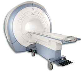
A technologist specially trained in MRI will answer any questions that you have regarding your test before performing your exam. Once you are positioned on the table, the table you are on will begin to move through the circular opening in the machine. The scan is painless.
A technologist will be just outside of the exam room during the procedure, at a viewing window. You will never be left alone. If indicated, you will follow simple Pre-Recorded directions through a speaker, telling you when to hold your breath and when to breathe so that your study gets the best results.
How long will my MRI take?
Allow 30 minutes to one hour for completion of your exam. Please also note this facility's ER can get busy which may cause a slight delay to scheduled patients. Emergencies do occur. For sedated children, there will be a recovery time in addition to this.
What will happen following my scan?
You may resume your activities unless instructed differently by your provider. You should feel no ill effects from your MRI scan. If you have been given IV contrast, we suggest drinking several glasses of water throughout the rest of the day, voiding often.
How do I find out the results of my MRI scan?
Your exam will be interpreted by a radiologist, dictated, than put in our computer system. Your provider will receive your results in about a week. If the radiologist finds something that needs immediate attention, the results will be communicated to your provider right away.
What if my child is having a MRI scan?
For a MRI scan to be successful, the patient is required to remain still during the exam. For this reason, some children may need to be sedated. We try to give the child a chance without sedation first for most non contrast studies. Parents may be allowed to accompany children into the MRI suite but will have to be screened prior his or her exam.
What types of exams are offered?
We have a variety of exams we do at BJACH. We can turn a simple scan of a head into a 3D image with our workstations. We do specialized studies for Surgery, Urology, EENT, Orthopedics and Emergency Room Trauma. These studies include:
- For studies of the head: Head, orbits, facial, sinuses, temporal bones, and 3D imaging.
- For studies of the neck: C-spine and soft tissue neck to include venous and arterial imaging.
- For studies of the chest: routine chest, thoracic dissection and follow up nodules.
- For studies of the Abdomen: Abdomen and Pelvis, Liver, Pancreatitis, KUB, Hematuria, Angio, Abdomen/Pelvis with runoff, and Abdominal Aortic Aneurysm.
- Other studies include: feet, ankles, knees, wrist, hands, elbows and shoulders.
Though your study may not be listed above, MRI provides an unparalleled view inside your body. It has become the preferred procedure for diagnosing a large number of potential problems in many different parts of the body.